Inside the Slow Progression of Glaucoma


Glaucoma is one of the leading causes of irreversible blindness worldwide, and among its many forms, chronic open-angle glaucoma stands as the most common and insidious. It progresses slowly, often without noticeable symptoms in its early stages, which is why it is frequently referred to as the “silent thief of sight.” Understanding how this condition develops, how it affects vision, and how it can be treated is crucial for both prevention and long-term management.
The Nature of Chronic Open-Angle Glaucoma
Chronic open-angle glaucoma is a long-term form of glaucoma in which the optic nerve — responsible for carrying visual information from the eye to the brain — suffers gradual damage. This damage occurs most commonly due to elevated intraocular pressure, although some individuals develop the condition even with normal pressure levels.
In the healthy eye, a clear fluid called aqueous humor circulates through a chamber behind the cornea and drains through a mesh-like structure known as the trabecular meshwork, located in the angle where the cornea meets the iris. In chronic open-angle glaucoma, this drainage system becomes less efficient. It does not close abruptly; instead, the resistance increases subtly and progressively. The eye continues to produce fluid, but the fluid drains out more slowly, causing the internal pressure of the eye to rise.
Over time, even moderately elevated pressure can harm the fragile fibers of the optic nerve. The process is gradual, typically occurring over several years. This slow progression is part of what makes chronic open-angle glaucoma so dangerous — patients rarely notice the change until significant vision loss has already occurred.
Why Early Detection Is Difficult
One of the defining characteristics of chronic open-angle glaucoma is the absence of early warning signs. The disease primarily affects peripheral vision at first, but the human brain is remarkably adept at compensating for mild visual field defects. Both eyes also work together, so a defect in one eye can be masked by normal vision in the other.
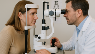
Because central vision remains intact until the disease is advanced, basic tasks such as reading or recognizing faces often feel completely normal. The gradual loss is subtle, leaving patients unaware of any problem. Routine eye examinations therefore play a pivotal role in detection. During an exam, ophthalmologists measure intraocular pressure, evaluate the optic nerve for signs of damage, and test the visual field for areas of reduced sensitivity.
Once vision is lost to glaucoma, it cannot be restored. For this reason, early diagnosis — before noticeable symptoms appear — is the most effective way to prevent severe impairment.
Recognizing the Symptoms as the Disease Advances
While early stages are silent, more advanced chronic open-angle glaucoma does create symptoms. These usually revolve around progressive narrowing of the visual field. Patients may notice trouble seeing objects out of the corner of their eye, difficulty navigating in dim light, or a general sensation that their environment feels “tunnel-like.” Driving, especially at night or in heavy traffic, becomes more challenging as peripheral awareness declines.
Some individuals experience mild eye discomfort or headaches, but these signs are non-specific and often dismissed. Unlike acute forms of glaucoma, chronic open-angle glaucoma does not create sudden pain, redness, or abrupt vision loss. The lack of dramatic symptoms is partly why the condition advances so far before being diagnosed.
How Chronic Open-Angle Glaucoma Differs From Angle-Closure Glaucoma
Although several types of glaucoma exist, the two major categories — open-angle and angle-closure — are fundamentally different. Understanding these differences helps clarify how chronic open-angle glaucoma behaves and why its treatment approach is distinct.
Open-angle glaucoma involves a drainage angle that remains anatomically open but functionally impaired. The trabecular meshwork is accessible to the circulating fluid, yet the meshwork itself fails to drain effectively. This dysfunction develops slowly, and pressure increases gradually.
In angle-closure glaucoma, the drainage angle becomes physically blocked, often by the iris shifting forward. This blockage prevents fluid from reaching the trabecular meshwork at all. The resulting pressure rise is rapid and dramatic, causing intense pain, blurred vision, nausea, halos around lights, and sometimes sudden visual loss. It is an ophthalmic emergency requiring immediate intervention.
Chronic open-angle glaucoma is therefore distinguished by slow progression, open but inefficient drainage, and long periods without symptoms. Angle-closure glaucoma, in comparison, is acute, painful, and often severe from the outset. The treatment strategies also differ, with angle-closure disease often requiring laser or surgical procedures to open or widen the angle.
Approaches to Treating Chronic Open-Angle Glaucoma
Treatment focuses on lowering intraocular pressure to slow or halt further damage to the optic nerve. While the lost vision cannot be reversed, pressure reduction can significantly reduce the risk of additional loss. Most patients begin with medicated eye drops that either decrease the production of aqueous humor or improve its outflow. Several classes of drops exist, including prostaglandin analogs, beta-blockers, carbonic anhydrase inhibitors, and alpha agonists.
If eye drops are insufficient or poorly tolerated, oral medications, laser therapy, or surgical procedures may be considered. Laser trabeculoplasty is commonly used to improve drainage through the trabecular meshwork. In some patients, this laser procedure can postpone or eliminate the need for long-term medications.
Surgical options such as trabeculectomy or minimally invasive glaucoma surgeries (MIGS) create new drainage pathways or enhance existing ones. The choice of treatment depends on factors such as the severity of disease, patient age, energy level of the optic nerve, and response to previous therapies.
The Role of Diamox in Managing Intraocular Pressure
Among the systemic medications used to treat glaucoma, Diamox (acetazolamide) holds particular importance. Diamox is a carbonic anhydrase inhibitor that reduces the production of aqueous humor inside the eye. While eye drops often target fluid production or drainage locally, Diamox works systemically, affecting the eye through the bloodstream.
Diamox is typically used in situations where rapid pressure reduction is needed or when topical medications have not provided adequate control. Because chronic open-angle glaucoma usually progresses gradually, Diamox is not always a first-line long-term therapy. However, it becomes extremely valuable when pressure rises beyond the safe range despite topical therapy, or when other medications are contraindicated.
Some patients use Diamox temporarily during acute spikes in pressure or as a bridge therapy while awaiting laser or surgical treatment. In more advanced cases, Diamox may be combined with multiple eye drops to achieve acceptable control.
Although effective, Diamox can cause side effects such as tingling in the fingers and toes, fatigue, altered taste sensation, frequent urination, and metabolic acidosis. Therefore, its use is carefully monitored. It remains an essential option in the broader treatment landscape, ensuring that pressure can be managed even when first-line therapies are insufficient.
Long-Term Management and Lifestyle Considerations
Chronic open-angle glaucoma is a lifelong condition that requires continuous monitoring. Even after treatment begins, the optic nerve may continue to weaken if pressure is not adequately controlled. Regular follow-up appointments, visual field tests, optic nerve imaging, and pressure measurements help ensure that therapy remains effective.
Patients also benefit from lifestyle habits that support ocular and cardiovascular health. Regular exercise, good blood pressure control, avoidance of smoking, and maintaining a healthy weight all contribute to a healthier optic nerve environment. Some studies suggest that diets rich in leafy greens, antioxidants, and omega-3 fatty acids may offer additional protective benefits, although these factors cannot replace medical treatment.
Preventive eye care is especially important for individuals with a family history of glaucoma, African or Hispanic ancestry, diabetes, or high myopia. These groups are at increased risk, making early screening crucial.
The Future of Glaucoma Treatment
Advancements in imaging technology, surgical techniques, and pharmacology continue to improve outcomes for patients with chronic open-angle glaucoma. New classes of medications, innovative minimally invasive surgical procedures, and genetic research are expanding the list of available tools. With earlier detection and more refined treatments, the long-term prognosis for many patients is improving.
Nevertheless, the fundamental principles remain unchanged: glaucoma must be diagnosed early, treated consistently, and monitored carefully. Delayed diagnosis still leads to preventable blindness in many parts of the world.
Protecting Vision Through Awareness and Treatment
Chronic open-angle glaucoma is both common and serious, but it is also manageable when detected early. By understanding how it develops, recognizing its progression, distinguishing it from other forms of glaucoma, and using medications such as Diamox appropriately, patients and clinicians can work together to preserve vision for as long as possible. With consistent care and modern treatment options, most people with chronic open-angle glaucoma can maintain functional vision throughout their lives.
Drug Description Sources: U.S. National Library of Medicine, Drugs.com, WebMD, Mayo Clinic, RxList.
Reviewed and Referenced By:
Dr. Robert N. Weinreb, MD Chair and Distinguished Professor of Ophthalmology at the Shiley Eye Institute, University of California San Diego. A world-leading authority on glaucoma pathophysiology and optic nerve damage, his research provides foundational insight into the progression and early detection of chronic open-angle glaucoma.
Dr. Teresa C. Chen, MD Associate Professor of Ophthalmology at Harvard Medical School and attending ophthalmologist at Massachusetts Eye and Ear. Widely cited on EyeWiki and Medscape, her clinical reviews focus on glaucoma diagnostics, surgical approaches, and long-term visual field preservation.
Dr. Anne L. Coleman, MD, PhD Professor of Ophthalmology at UCLA Stein Eye Institute. Her work with the National Eye Institute explores population risk factors, glaucoma-related visual disability, and the effects of early intervention on disease progression.
Dr. Paul J. Foster, MD, PhD Professor of Ophthalmic Epidemiology and Glaucoma Studies at University College London. A principal investigator for several major glaucoma trials, his research distinguishes open-angle from angle-closure mechanisms and provides evidence-based guidance for global glaucoma care.
(Updated at Nov 21 / 2025)