Neural tube: what it is, how it is formed, and associated diseases.
It is one of the most important elements of fetal development, and from it arises the nervous system.
The complexity of our nervous system, the fundamental system that connects and governs all the processes of our organism, is something that continues to amaze the many researchers and experts who study it every day. But one fact must be taken into account, and that is that although when we think of a nervous system we generally think of a mature structure, it is necessary that a series of processes take place since we are little more than an accumulation of cells to reach a mature nervous system.
Throughout embryonic and fetal development, a series of events will take place that will trigger the formation of the so-called neural tube, which in turn will develop during gestation until the structures of the nervous system to generate the structures of the human nervous system
What is the neural tube?
The neural tube is known as the the structure that is formed during gestation and that is the immediate ancestor of the nervous system, being its closure and evolution the one that will end up generating the different structures that are part of it.It is its closure and evolution that will eventually generate the different structures that are part of it. Specifically, we are talking about the brain and the spinal cord, being others such as the peripheral nervous system formed by the neural crests.
Technically, the process in which the neural tube is generated and closes would start from the third week of gestation and should finish closing approximately around the twenty-eighth day. It should be noted that it is essential for the tube to close so that the spine and skull can protect the nerves and brain and so that they can form. This closure is usually successful in most births, but sometimes the tube fails to close, which can lead to various neural tube defects.
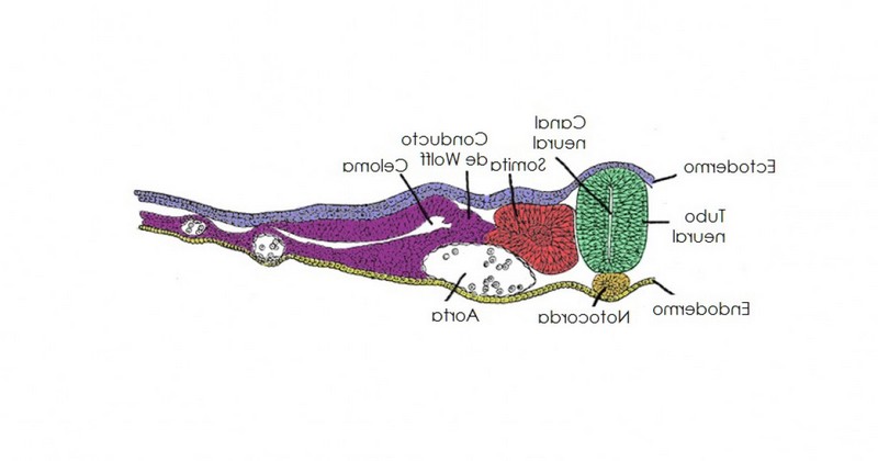
Neurulation: formation and development of the neural tube
The neural tube is produced through a process known as neurulationin which the notochord and the mesoderm as a whole lead the ectoderm to differentiate into the neuroectoderm. This thickens and eventually detaches from the cell sheet, forming the neural plate.
This plate will proceed to stretch rostrocaudally, in such a way that it will generate folds, which will grow with the development of the fetus. With time, a sinking of the central part occurs, generating a channel whose walls will close in on themselves until a tube-shaped structure is generated: the neural tube. This tube begins to close on itself in the middle part, advancing towards the ends. In this process the neural crests are also separated and detached from the tube.which will end up generating the autonomic nervous system and different organs and tissues of the different body systems.
Initially the tube will be open at its ends, forming the rostral and caudal neuropores, but from the fourth week they begin to close. This closing and the development of the tube will generate various dilations in its rostral-cranial part, which in the future will form the different parts of the brain. Generally, the rostral end closes first, around day 25, while the causal end usually closes around day 27.
There is a second process of neurulation, the so-called secondary neurulation, in which the part of the neural tube corresponding to the spinal column is formed and at the same time hollowed out in such a way that the internal cavity of the neural tube is emptied, the internal cavity of the neural tube is emptied, generating a separation between epithelium and mesenchymal cells (which will form the (which will form the medullary cord). In the medulla we find that motor neurons appear in the ventral part, while sensory neurons appear in the dorsal part of the medulla.
Formation of the different brain regions
Throughout the formation and development of the neural tube, the structures that form part of our adult nervous system will be produced. The cells of the neural tube, once closed, begin to divide and generate different layers and structures. It will be in the anterior or cranial face of the tube where the brain will appear.
During the fourth week of gestation, prosencephalon, midbrain and rhombencephalon can be observed.. During the fifth week, the first and the third divide, forming telencephalon and diencephalon in the first and metencephalon and myelencephalon in the second. In a relatively fast way, the structure changes in a heterogeneous way, growing the different structures (being the telencephalon, the proper part of the cortex, the one that develops the most).
It is important to bear in mind that not only the wall of the neural tube is important, but also the hollows and empty spaces inside it: they will eventually form the ventricles and the set of structures through which the cerebrospinal fluid will circulate, without which the brain could not function properly.
Neurulation defects
The process of neurulation, in which the structure of the nervous system is formed, is fundamental to the human being. However, alterations and malformations sometimes alterations and malformations can occur, which can have more or less severe consequences that can have more or less severe consequences on the development and survival of the fetus. Among them, some of the best known are the following.
1. Spina bifida
One of the most common and well-known neural tube defects is spina bifida. known neural tube defects is spina bifida. This alteration supposes the existence of some type of problem that prevents a part of the neural tube from closing completely, something that can have effects of varying severity because the nerves and the spinal cord cannot be properly protected by the spinal column.
Within this type of alterations we can find subjects whose alteration is not visible (hidden), although they may have holes or protuberances in the back, and others who have a directly perceptible hole (cystic or open). The closer it is to the brain, the more serious the possible nerve injuries may be.
2. Anencephaly
Another of the best known neural tube defects and alterations is anencephaly. In this case, we observe that the caudal part of the neural tube has not closed completely. This alteration is usually incompatible with life, and it is not uncommon for babies to miscarry or have a very short life expectancy after birth.. However, in some cases survival is more prolonged. Anencephalic subjects cannot perform complex cognitive and sensory functions, having no awareness of the environment or of themselves and in most cases not being able to perceive (although they may have reflexes).
3. Encephalocele
Alteration produced by problems during the closure of the rostral end of the neural tube. Equivalent to spina bifida but in the skull, it involves the existence of a protrusion of part of the contents of the brain to the outside of the skull, generally presenting a kind of sac or lump in the head with such contents.The child usually presents a kind of sac or lump in the head with such contents. In most cases cognitive alterations are generated, and it is not infrequent the death of the minor during the fetal development.
4. Chiari malformation
It is common that the presence of alterations in the development and closure of the neural tube generate the so-called Chiari malformations, which consist of a protrusion of part of the cerebellum or part of the brain into the spinal canal, being displaced by some type of structural malformation of the skull or brain. In other words, part of the content of the brain invades and occupies the spinal canal. It may not generate symptoms, but it can also generate pain, balance, vision and coordination problems and paresthesias.
Bibliographic references
- López, N. (2012) Developmental biology. Workbook, McGraw-Hill Education.
(Updated at Apr 13 / 2024)