Tricipital reflex: what it is, physiology, and how it is analyzed.
These are the characteristics of the tricipital reflex and its relation to health examinations.
The neurological examination is, together with the anamnesis, the basis for the diagnosis of nervous system pathologies. Although many complex tests can be performed to evaluate Muscle tone and the intensity of contractions, one of the first steps in the suspicion of a problem at the nervous system level is always to quantify osteotendinous reflexes.
These reflexes are fast and involuntary, as they do not pass through the brain and are processed at the level of the spinal cord. Due to their variety and knowledge about them, it is possible to detect very accurately if some kind of damage has occurred in the spinal cord and, if so, it is also possible to locate the injury very accurately. When osteotendinous reflexes are quantified, the strength and speed of the contraction is measured, as well as the symmetry (or lack thereof) of the response and the homogeneity of reaction between different parts of the body.
In this type of test it is not so important to put a number to each reflex from 1 to 5, but to quantify the variability between the different sections and neural pathways in the patient. Based on these very interesting premises, we tell you all about the tricipital reflex.
What are the osteotendinous reflexes?
The tricipital reflex is a type of osteotendinous reflex, so before we look at it, we must lay some groundwork as far as medical terminology is concerned.
The osteotendinous reflexes are a type of medullary reflex, whose route of action is extremely circumscribed and very fast.. This type of action does not pass directly through the brain, hence the rapidity of the association between stimulus and response.
When a force is applied to the critical point of the muscle environment, the muscle involuntarily lengthens. The neuromuscular spindle (sensory receptors within the muscle) then sends the mechanical stress signal to an afferent neuron, which in turn contacts the nerve center. The dorsal root ganglia pick up this stimulus, which is interpreted directly in the gray matter of the spinal cord. Finally, the motor neuron axons leave the medulla and send the muscle contraction signal.
As you can see, we are dealing with a very circumscribed closed circuit: spindle-neuron afferent-spinal cord-motor neuron. Because the information does not pass through the brain the information does not pass through the brain and is always interpreted on the same levelIn this way, it is possible to detect neurological faults very accurately by quantifying the osteotendinous reflexes. The most important are the following:
- Bicipital: it is investigated on the inner side of the elbow.
- Tricipital: consists of the percussion of the triceps.
- Stylo-radial: the styloid process of the radius is percussed, where the tendon of the supinator longus is inserted.
- Ulnar-pronator: percussion is performed at the level of the ulnar styloid.
- Rotulian: the most famous of all. The patellar tendon is percussed, which generates an involuntary lifting of the leg.
- Achilles: the Achilles tendon, which connects the calf muscle at the back of the leg to the heel bone, is percussed.
What is the tricipital reflex?
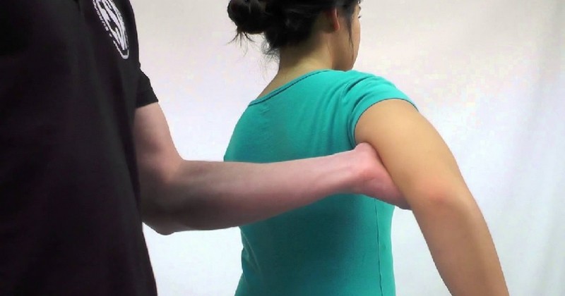
As we have said, the tricipital reflex is a type of osteotendinous reflex, and in turn myotatic, since the synaptic connection is made at the level of the spinal cord (and not the brain). To assess its functionality, a force is applied to the triceps tendon, located above the elbow (at the olecranon).. With this reflex, the nerve roots C6, C7 (predominant) and C8, or in other words, the integrity of the musculocutaneous nerve, are brought into play.
To perform this test, the patient's forearm should be held (ideally placed on the thigh), with the arm in a position midway between flexion and extension. Once the desired position has been reached, the triceps tendon should be located and percussed at its base of insertion.
When this sudden force is applied, the forearm is expected to extend rapidly.. If the absence of movement is absolute (areflexia), myopathy, neuropathy, spondylosis and other clinical entities of a neuromuscular nature are suspected.
How is it tested in patients?
In order to homogenize such a subjective test, the National Institute of Neurological Disorders and Stroke devised a numerical scale applicable in all cases. Once percussion is produced in the tendon of interest, the response is quantified based on the following parameters:
- 0: no response at all, considered as areflexia.
- 1: reflex is present, but very weak and less pronounced than normal. It includes a trace response or, alternatively, a response that appears with reinforcement.
- 2: reflex occurs, but is below the expected "half" or normal range.
- 3: the reflex occurs, above the expected "half" or normal range.
- 4: reflex occurs, more than normal. Clonus, involuntary and rhythmic contractions of the muscle environment after stimulation, may occur.
- 5: Not always used, but this category may reflect sustained clonus.
Depending on the other responses, a tricipital reflex between 2 and 3 can be considered as "normal", as long as it occurs in the same way in both body planes (left and right arm). (left and right arm). A value of 0 is conceived as areflexia, while a value of 4-5 is hyperreflexia.
In addition, it should be noted that each of these values can be further circumscribed with a (+) or (-), reflecting that the patient's picture falls between two of the figures. As you can imagine, a 3+ and a 4- can be the same for two different evaluators, so we repeat that it is not so important the sign and the number as the homogeneity between results within the same patient.
The interpretation of the results
An areflexia may evidence damage at the level of a particular nerve pathway or, alternatively, an abnormality in the spinal cord or the entire nervous system. and general condition of the patient. Without going any further, some of the osteotendinous reflexes are better predictors of diabetes-derived neuropathy than many other tests and subjective symptoms of the patient.
Hyperreflexia may indicate damage to the upper motor neurons, while hyporeflexia or areflexia usually indicates damage to the lower motor neurons. In general, a 1+ to 3+ range is considered to be within normal if the response is symmetrical. In any case, even an absent reflex may be considered normal in some patients, if it is not accompaniedHowever, even an absent reflex may be considered normal in some patients, if it is not accompanied by other symptoms and conditions that would suggest a problem at the neurological level.
Today, peripheral neuropathies are the most common cause of absent reflexes in general society. The triggers for this condition are varied: diabetes, alcoholism, amyloidosis, uremia, vitamin deficiencies, pernicious anemia, remote cancer, presence of toxins in the body and many other etiological agents. Once an abnormality in the tricipital reflex (or any osteotendinous reflex) is detected, further tests are needed until the patient's problem is found.
Summary and final notes
As you have seen, the tricipital reflex (and the osteotendinous reflexes in general) is essential for clinical semiology, especially when detecting a neuropathy of the peripheral nervous system. These reflexes are extremely important in the medical field, because as they do not "pass" through the brain, damage can be clearly detected in a very specific environment in the section of the spinal cord involved.
Thus, reflexes are very useful in detecting pathologies, but they must be accompanied by a series of accessory tests to confirm or rule out a diagnosis. The osteotendinous reflexes are the first step to suspect a disease, but they never make up by themselves the complete diagnosis.
(Updated at Apr 14 / 2024)