Ischial (sciatic) nerve: anatomy, functions and pathologies

This nerve is the one that, if affected, causes the much-feared sciatica.
We have all heard of (or have suffered in our own flesh) the discomfort caused by a disorder such as sciatica.
The main cause of this characteristic pain is the compression of the ischial nerve, which causes intense pain and numbness in the limbs. It is precisely this important nerve that we will discuss in this article.
We explain what it is and where it is located, as well as its main functions.. We will also discuss the various disorders associated with ischial nerve injury.
- Recommended article: "The 11 main diseases of the spine".
Ischial nerve: definition, structure and location.
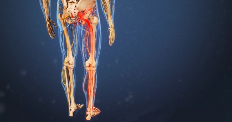
The ischial nerve, also called the sciatic nerve, is the largest and longest peripheral nerve in humans and other vertebrates. and other vertebrate animals. It begins in the pelvis, at the bottom of the sacral plexus, formed by the anterior roots of several spinal nerves, and continues through the hip joint, down the leg.
In humans, the ischial nerve is formed from the L4 and S3 segments of the sacral plexus, whose fibers unite to form a single nerve in front of the piriformis muscle. The nerve then passes under this Muscle and through the greater sciatic foramen, exiting the pelvis.
From there it travels down the posterior thigh to the popliteal fossa (colloquially known as the "hamstring"). The nerve passes through the posterior compartment of the thigh behind the adductor magnus muscle, in front of the long head of the biceps femoris muscle.
The ischial nerve, in the lower thigh area and above the knee (posteriorly), divides into two nerves: the tibial nerve, which continues its descending path towards the feet and is responsible for innervating the heel and sole; and the peroneal nerve, which runs laterally along the outside of the knee and up to the upper foot area.
As we will see later, this nerve provides the connection to the nervous system for almost all of the skin of the leg, the muscles of the back of the foot, and the muscles of the lower leg.The muscles of the back of the thigh and those of the leg and foot. Next, we will see what functions this important nerve is responsible for.
Functions
The ischial nerve is the nerve that allows movement, reflexes, motor and sensory functions and strength to the leg, thigh, knee, calf, ankle, ankle and lower leg.calf, ankle, toes and feet. Specifically, it serves as a connection between the spinal cord and the outer thigh, the hamstring muscles found in the back of the thigh, and the muscles of the lower leg and feet.
Although the ischial nerve passes through the gluteal region, it does not innervate any muscles there. However, it does directly innervate muscles in the posterior compartment of the thigh and the hamstring portion of the adductor magnus muscle. Through its two terminal branches, it innervates the calf muscles and some muscles of the foot, as well as those of the anterior and lateral leg, and some other intrinsic muscles of the foot.
On the other hand, although the ischial nerve does not have cutaneous functions per se, it does provide indirect sensory innervation through its terminal branches by innervating the posterolateral anterolateral sides of the leg and the sole of the foot, as well as the lateral part of the leg and dorsal area of the foot.
Related disorders: sciatica
Sciatica is the result of damage or injury to the ischiatic nerve and is characterized by a sensation that may manifest with symptoms of moderate to severe pain in the back, buttocks and legs. Weakness or numbness may also occur in these areas of the body. Typically, the person experiences pain that flows from the lower back, through the buttocks and into the lower extremities.
Symptoms are usually made worse by sudden movement (e.g. getting out of bed), by certain positions (e.g. sitting for a long time) or when performing physical exercise with weights (e.g. moving a piece of furniture or picking up a bag). The most common causes of sciatica include the following:
1. Herniated discs
The vertebrae are separated by pieces of cartilagewhich is filled with a thick, transparent material that ensures flexibility and cushioning when we move. Herniated discs occur when that first layer of cartilage is torn.
The substance inside can compress the ischial nerve, resulting in pain and numbness in the lower extremities. It is estimated that between 1 and 5 percent of the population will suffer back pain caused by a herniated disc at some point in their lives.
2. Spinal stenosis
Spinal stenosis, also called lumbar spinal stenosis, is characterized by abnormal narrowing of the lower spinal canal.. This narrowing puts pressure on the spinal cord and its sciatic nerve roots. Symptoms that may be experienced are: weakness in the legs and arms, lower back pain when walking or standing, numbness in the legs or buttocks, and balance problems.
3. Spondylolisthesis
Spondylolisthesis is one of the associated conditions of degenerative disc disorder.. When one vertebra extends forward over another, the extended spinal bone can pinch the nerves that form its ischial nerve.
Although it is a painful condition, it is treatable in most cases. Symptoms include: stiffness in the back and legs, persistent low back pain, thigh pain, and tightness of the hamstrings and gluteal muscles.
4. Piriformis syndrome
Piriformis syndrome is a rare neuromuscular disorder in which the piriformis muscle contracts or tightens involuntarily, causing sciatica. This muscle is the muscle that connects the lower spine to the thigh bones. When tightened, it can put pressure on the ischial nerve..
Clinical features of the syndrome include radicular pain, muscle numbness and weakness, and tenderness in the buttocks. Occasionally, the pain may be exacerbated by internal rotation of the lower extremity of the hip.
The usual treatment is usually surgical, with the aim of releasing the piriformis muscle; or non-surgical, with the injection of corticosteroid drugs, the application of analgesic medication and physiotherapy.
Bibliographic references:
-
Cardinali, D.P. (2000). Manual of neurophysiology. Madrid: Ediciones Díaz de Santos.
-
Olmarker, K., & Rydevik, B. (1991). Pathophysiology of sciatica. The Orthopedic clinics of North America, 22(2), 223-234.
-
Sobotta, J. (2006). Atlas de anatomia humana (Vol. 2). Ed. Médica Panamericana.
(Updated at Apr 13 / 2024)