Acute Leukaemia: Types, Symptoms and How It Is Treated

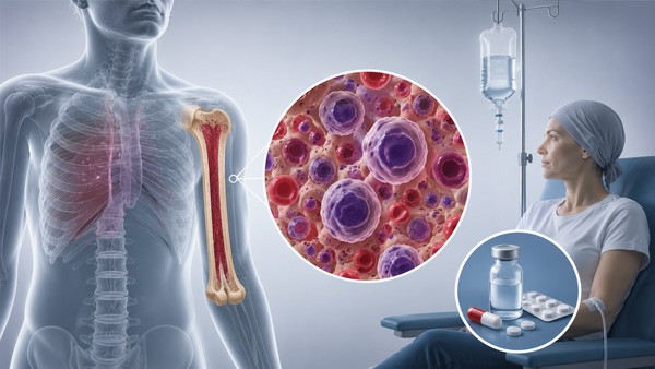

Acute leukaemia is one of the few cancers that behaves like an emergency. Where most malignancies develop over months or years, acute leukaemia can take a person from apparently well to critically ill in a matter of weeks — and, unusually among cancers, that speed cuts both ways: it is also among the diseases where prompt intensive treatment produces cure rather than control.
This guide covers what the disease is, how the two main types differ, which presentations are medical emergencies, how diagnosis is established, what modern treatment involves, and what the realistic outlook is for different groups. It is written to help patients and families understand what is happening, not to guide treatment — acute leukaemia is managed in hospital by haematology teams from the day of diagnosis.
🚨 If this describes someone now: unexplained bruising or bleeding, fever with a recent abnormal blood count, extreme fatigue with pallor and breathlessness, or a blood test showing very high or very low white cells — that person needs assessment today, not an appointment next week. Acute leukaemia is diagnosed and treated as an emergency, and delay of days can matter.
🩸 What is happening in the bone marrow
Blood cells are produced in the bone marrow from stem cells that mature through a series of stages. In acute leukaemia, a cell early in that sequence acquires genetic changes that lock it in an immature state — a blast — while removing the normal signals that stop it dividing. The blasts multiply, fill the marrow, and crowd out production of everything else.
That crowding explains the entire clinical picture. Falling red cells produce anaemia: fatigue, pallor, breathlessness. Falling normal white cells leave the person unable to fight infection. Falling platelets produce bruising and bleeding. The blasts themselves spill into the bloodstream and can infiltrate the liver, spleen, lymph nodes, gums, skin and central nervous system.
The word acute is not a description of severity but of cell maturity: acute leukaemias involve immature blasts and progress rapidly, while chronic leukaemias involve more mature cells and evolve over years. Diagnosis conventionally requires 20% or more blasts in the marrow or blood, though certain genetic abnormalities establish the diagnosis at lower counts.
🔠 The two main types
Acute leukaemia divides according to which cell line the malignant clone arose from, and the distinction determines treatment entirely.
| Feature | Acute lymphoblastic leukaemia | Acute myeloid leukaemia |
|---|---|---|
| Cell of origin | Lymphoid precursors | Myeloid precursors |
| Typical age | Peaks in early childhood; also occurs in adults | Predominantly older adults, median age around 68 |
| Central nervous system involvement | Common enough that prophylaxis is routine | Less frequent |
| Treatment duration | Two to three years including maintenance | Intensive blocks over months, without long maintenance |
| Outlook in children | Very good — survival above 90% in modern protocols | Less favourable, though improving |
| Outlook in adults | Intermediate, improving with newer agents | Highly variable, driven by genetics and fitness |
Within each type, genetic subgroups matter more than the broad category. Some abnormalities predict excellent response to standard treatment; others indicate a need for transplantation. This is why treatment is not decided until the genetic workup returns, except where the situation is too urgent to wait.
📋 How it presents
Symptoms develop over days to weeks and reflect marrow failure rather than a lump or a mass.
- Fatigue, pallor and breathlessness on minimal exertion — from anaemia, frequently the first thing noticed.
- Fever and recurrent or unusual infections — from the absence of functioning neutrophils.
- Easy bruising, petechiae, nosebleeds, bleeding gums, heavy periods — from low platelets.
- Bone and joint pain, particularly in children, from marrow expansion — sometimes mistaken for growing pains or arthritis.
- Swollen lymph nodes, enlarged liver or spleen, more typical of the lymphoblastic type.
- Weight loss, night sweats, loss of appetite.
- Gum swelling or skin infiltration, characteristic of certain myeloid subtypes.
Individually each of these has commoner explanations, which is why the diagnosis is frequently made when a blood count ordered for fatigue returns markedly abnormal. That full blood count is the single most important test in the whole pathway, and it is cheap and universally available.
🚨 The four emergencies
Several complications kill quickly and are managed before or alongside the leukaemia itself.
| Emergency | What it is | Why it is urgent |
|---|---|---|
| Febrile neutropenia | Fever in a person with almost no neutrophils | Sepsis can progress within hours; antibiotics are given immediately, before results |
| Acute promyelocytic leukaemia | A specific myeloid subtype with severe coagulopathy | High early death rate from haemorrhage; specific treatment starts on suspicion alone |
| Hyperleukocytosis and leukostasis | Extremely high blast counts clogging small vessels | Causes breathlessness, confusion and stroke-like events; needs urgent cytoreduction |
| Tumour lysis syndrome | Massive cell breakdown releasing potassium, phosphate and uric acid | Causes arrhythmia and kidney failure; prevented with hydration and specific drugs |
⚠️ Acute promyelocytic leukaemia deserves separate emphasis because it is the clearest example of why urgency matters. Untreated, it kills rapidly through bleeding. Recognised and treated promptly with differentiating agents rather than conventional chemotherapy alone, it has become one of the most curable adult leukaemias — with cure rates above 90% in suitable patients. The entire difference is made in the first hours and days.
Febrile neutropenia is the emergency families most need to recognise, because it happens at home between hospital visits. A temperature of 38 degrees or above in someone receiving chemotherapy is an emergency requiring immediate attendance, regardless of how well the person otherwise appears. Waiting to see whether it settles is the mistake that costs lives.
🔬 How the diagnosis is established
The pathway moves quickly and involves several layers, each answering a different question.
Full blood count and blood film raise the suspicion, showing anaemia, abnormal white cell counts, low platelets and circulating blasts. Bone marrow aspiration and trephine biopsy confirm it, usually taken from the back of the pelvis under local anaesthetic.
The marrow sample then goes through three separate analyses. Immunophenotyping by flow cytometry identifies which cell line the blasts belong to, separating lymphoblastic from myeloid disease. Cytogenetics examines chromosomes for translocations and other abnormalities that define prognostic groups. Molecular testing identifies specific mutations, several of which now have drugs targeting them directly.
Additional tests include a lumbar puncture to check for central nervous system involvement, particularly in lymphoblastic disease; imaging where organ infiltration is suspected; and baseline cardiac and organ function testing before treatment that can affect the heart, liver and kidneys.
💊 Treating acute lymphoblastic leukaemia
Treatment is prolonged and structured in phases, and the structure is what produces the high cure rates in children.
Induction aims to clear detectable disease and restore normal marrow function, typically over four to six weeks with a combination of agents including corticosteroids, vincristine and asparaginase. Consolidation follows, using intensive blocks to eliminate residual disease. Maintenance then continues for two to three years with lower-intensity oral treatment — commonly mercaptopurine with methotrexate — because shortening this phase increases relapse.
Throughout, central nervous system prophylaxis is given, since leukaemic cells hide in the CNS where most chemotherapy penetrates poorly. This is delivered as intrathecal chemotherapy injected into the spinal fluid, and it is one of the innovations that transformed outcomes.
Where the Philadelphia chromosome is present, a tyrosine kinase inhibitor such as imatinib or dasatinib is added, converting a formerly poor-prognosis subgroup into one that responds well.
🧬 Treating acute myeloid leukaemia
Historically, treatment meant intensive chemotherapy combining cytarabine with an anthracycline over seven days, followed by consolidation courses. That regimen remains standard for patients fit enough to tolerate it.
The major recent change concerns everyone else. Many patients with AML are elderly or have other illnesses that make intensive chemotherapy unsurvivable, and for decades those patients received little more than supportive care. The combination of venetoclax with a hypomethylating agent changed that substantially, producing meaningful remission rates in patients previously considered untreatable.
Alongside this, targeted drugs now address specific mutations: inhibitors for FLT3, for IDH1 and IDH2, and antibody-drug conjugates for CD33-positive disease. Molecular testing at diagnosis determines which apply, which is why treatment decisions wait for those results where the clinical situation permits.
📊 Measurable residual disease has become central. Modern techniques detect leukaemic cells at levels far below what a microscope can see, and the presence or absence of that residual disease after treatment predicts relapse better than any traditional marker. It increasingly determines who proceeds to transplantation and who does not.
🏥 Stem cell transplantation
Allogeneic transplantation replaces the patient marrow with stem cells from a donor, after chemotherapy or radiotherapy has eliminated the existing marrow. Its power comes partly from the conditioning treatment and partly from the donor immune cells recognising and attacking residual leukaemia.
It is reserved for patients whose disease carries high relapse risk, or who have relapsed and achieved a second remission, because it carries substantial risks of its own: graft-versus-host disease, serious infection during the period without immunity, and organ toxicity. The decision balances the risk of the leukaemia returning against the risk of the procedure, and it is one of the more difficult conversations in medicine.
🛡️ Supportive care, which does much of the saving
A substantial share of the improvement in survival over recent decades came not from better chemotherapy but from better support during it.
- Transfusion support — red cells for anaemia, platelets to prevent bleeding while the marrow recovers.
- Infection prevention and rapid treatment — antimicrobial prophylaxis, central line care, and immediate broad-spectrum antibiotics at the first fever.
- Growth factors to shorten the period of neutropenia in selected situations.
- Tumour lysis prevention with hydration and drugs that lower uric acid before treatment begins.
- Nutrition, mouth care and antiemetics, which determine whether a patient can complete the planned treatment.
- Fertility preservation, discussed before treatment starts where possible, since several agents affect future fertility.
- Psychological and social support for a diagnosis that arrives without warning and upends a family within days.
📈 What the outlook actually is
Prognosis varies more within acute leukaemia than between most other cancers, and averages mislead badly. Three factors dominate: the type, the genetic subgroup, and the age and fitness of the patient.
Childhood lymphoblastic leukaemia is among the great successes of modern medicine, with long-term survival above 90% in current protocols — a transformation from a uniformly fatal disease within living memory. Adult lymphoblastic leukaemia does less well, though immunotherapies including bispecific antibodies and CAR-T cell therapy have improved options in relapsed disease considerably.
Acute myeloid leukaemia remains harder. Younger patients with favourable genetics can be cured with chemotherapy alone; older patients with adverse genetics face a difficult outlook, though targeted agents and venetoclax-based combinations have improved what was previously available. Age matters here more than in almost any other malignancy, because it determines what treatment a person can survive.
✅ Two questions worth asking the haematology team early: what is the genetic subgroup and what does it mean for the plan? And is there a clinical trial for which this situation qualifies? Acute leukaemia is an area where trial participation frequently provides access to agents not otherwise available, and where the pace of change means today standard differs from that of five years ago.
🏷️ About medication and this catalogue
Acute leukaemia is treated with intravenous chemotherapy, targeted agents, transfusions and transplantation, delivered in hospital under specialist supervision. It cannot be treated with medicines ordered online, and no part of this article should be read as suggesting otherwise.
Two drugs held in the oncology category do appear in leukaemia protocols in specific roles — methotrexate features in maintenance and central nervous system treatment of lymphoblastic disease, and hydroxyurea is used to reduce very high white cell counts. Both are prescribed, dosed and monitored by a haematology team as part of a protocol, with regular blood testing; neither is something a patient starts independently.
❓ Frequently asked questions
What makes leukaemia acute rather than chronic?
The maturity of the abnormal cells rather than the severity of the illness. Acute leukaemia involves immature blasts that cannot function and multiply rapidly, producing symptoms within weeks. Chronic leukaemia involves more mature cells and evolves over years, sometimes without symptoms for a long period. Diagnosis of acute disease conventionally requires 20% or more blasts in marrow or blood.
What are the first symptoms?
They reflect bone marrow failure: fatigue, pallor and breathlessness from anaemia; fever and infections from lack of functioning white cells; easy bruising, petechiae and bleeding from low platelets. Bone pain is common in children. Each symptom has commoner explanations, which is why the diagnosis is often made when a full blood count ordered for fatigue returns markedly abnormal.
Why is it treated as an emergency?
Because it progresses over days to weeks rather than months, and because several complications can kill quickly — sepsis in a person without neutrophils, bleeding from severe coagulopathy in one myeloid subtype, vessel obstruction from extremely high blast counts, and metabolic collapse when cells break down. Treatment frequently begins the same week the diagnosis is suspected.
What should we do if there is a fever during chemotherapy?
Attend hospital immediately, without waiting to see whether it settles. A temperature of 38 degrees or above in someone with low neutrophils is a medical emergency, because infection can progress to sepsis within hours. Antibiotics are given promptly, before culture results return. This is the single most important thing for families to know between hospital visits.
How is the diagnosis confirmed?
By bone marrow aspiration and biopsy after an abnormal blood count. The sample undergoes immunophenotyping to identify the cell line, cytogenetic analysis for chromosomal abnormalities, and molecular testing for specific mutations. A lumbar puncture checks for central nervous system involvement. Those results determine the treatment plan, which is why they are awaited where the clinical situation allows.
Why does treatment for lymphoblastic leukaemia last years?
Because after intensive induction and consolidation, a prolonged maintenance phase of lower-intensity oral treatment is needed to prevent relapse. Shortening it increases the chance of the disease returning. Central nervous system prophylaxis is also given throughout, since leukaemic cells can shelter in the spinal fluid where most chemotherapy penetrates poorly.
What changed for older patients with myeloid leukaemia?
For decades, patients too frail for intensive chemotherapy received little beyond supportive care. Combinations of venetoclax with a hypomethylating agent changed that, producing meaningful remission rates in people previously considered untreatable. Targeted drugs for specific mutations, including FLT3 and IDH inhibitors, have added further options identified through molecular testing at diagnosis.
When is a stem cell transplant needed?
Generally when the genetic subgroup indicates high risk of relapse, or after relapse once a second remission is achieved. It offers the strongest chance of cure in those situations but carries substantial risks: graft-versus-host disease, serious infection, and organ toxicity. The decision weighs the likelihood of the leukaemia returning against the risks of the procedure itself.
What is measurable residual disease?
Leukaemic cells detectable by sensitive laboratory techniques at levels far below what microscopy can see. Its presence after treatment predicts relapse better than traditional markers, and it increasingly guides decisions about whether to proceed to transplantation or to intensify therapy. A patient in apparent remission by microscope may still have detectable residual disease.
What is the survival rate?
It varies enormously and averages mislead. Childhood lymphoblastic leukaemia now has long-term survival above 90% in modern protocols. Adult lymphoblastic disease does less well, though immunotherapy has improved relapsed disease considerably. Myeloid leukaemia outcomes depend heavily on genetics and on whether the patient can tolerate intensive treatment, with age being the dominant practical factor.
Can acute leukaemia be treated with tablets bought online?
No. Treatment requires intravenous chemotherapy, transfusion support, management of life-threatening complications and frequently transplantation, all delivered in hospital under haematology supervision. Some oral drugs feature within protocols, but they are prescribed, dosed and monitored as part of a supervised plan with regular blood testing. Attempting to self-treat this disease is fatal.
Should fertility be discussed before treatment?
Yes, wherever the clinical urgency permits. Several agents used in these protocols affect future fertility, and options for preservation exist for both men and women but must be arranged before treatment begins. It is easily overlooked in the rush of an emergency diagnosis, which is why raising it early matters, particularly for younger patients.
📑 Sources and editorial
- World Health Organization classification of haematolymphoid tumours — diagnostic criteria and genetic subgroups
- NCCN and ESMO clinical practice guidelines for acute lymphoblastic and acute myeloid leukaemia
- Published trial evidence on venetoclax combinations, FLT3 and IDH inhibitors, and immunotherapy in relapsed disease
- Guidance on febrile neutropenia, tumour lysis syndrome and management of acute promyelocytic leukaemia
- Population survival data for childhood and adult acute leukaemia from national cancer registries
- RXshop Editorial Team — reviewed by Emily Chen, MD, MPH, Internal Medicine
Medical Disclaimer: The information in this article is for educational and informational purposes only and is not a substitute for professional medical advice, diagnosis, or treatment. Always seek guidance from a qualified healthcare provider with any questions you may have regarding a medical condition, and before starting, stopping or changing any medication.