Choriocarcinoma: What It Is and Why It Is Highly Curable

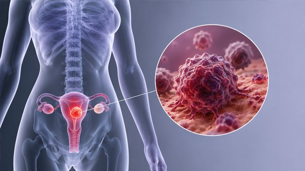

Choriocarcinoma is a rare cancer that arises from placental tissue, spreads early and aggressively to the lungs, brain and liver — and is cured in the great majority of women who develop it, including many with widespread metastases. That combination is almost unique in oncology, and it is worth stating at the outset, because a diagnosis with the word carcinoma in it rarely comes with this prognosis.
Two things make it possible. The tumour is exceptionally sensitive to chemotherapy, and it produces a hormone that can be measured in blood with great precision, giving clinicians a marker that tracks the disease more accurately than any scan. This guide covers what the condition is, how it presents, why that hormone matters so much, how treatment is selected, and what it means for future pregnancies.
🧬 The essential context: choriocarcinoma belongs to a family of conditions called gestational trophoblastic disease, which arise from the tissue that would normally form the placenta. Some members of that family are benign, some malignant. What they share is an origin in pregnancy tissue and a hormone marker that makes them unusually trackable.

🩸 What the disease actually is
During normal pregnancy, trophoblast cells invade the uterine wall to establish the placenta, and invasion is part of their ordinary function. In gestational trophoblastic disease, that tissue proliferates abnormally. Where the abnormal tissue becomes malignant and invasive, it is termed gestational trophoblastic neoplasia.
| Condition | Nature | What happens |
|---|---|---|
| Complete or partial hydatidiform mole | Benign | Abnormal pregnancy tissue; removed surgically, then monitored |
| Invasive mole | Malignant, locally invasive | Molar tissue penetrating the uterine wall |
| Choriocarcinoma | Malignant, metastasises early | Aggressive spread via bloodstream; highly chemosensitive |
| Placental site trophoblastic tumour | Malignant, rare | Slower growing, less chemosensitive — treated surgically |
| Epithelioid trophoblastic tumour | Malignant, very rare | Similar behaviour to the above; surgery is central |
Choriocarcinoma is uncommon — on the order of one case per tens of thousands of pregnancies in Western populations, with higher rates reported in parts of Asia and Latin America. Hydatidiform mole, the condition that precedes many cases, occurs considerably more often, at roughly one per thousand pregnancies.
📅 Which pregnancies it follows
Choriocarcinoma always arises from a pregnancy, but not necessarily a recent or a normal one. Roughly half of cases follow a hydatidiform mole; the remainder follow a normal-term delivery, a miscarriage or an ectopic pregnancy in approximately equal proportions.
That distribution has an important practical consequence. A woman who develops abnormal bleeding months after an entirely normal delivery is not usually suspected of having a cancer, and the diagnosis is delayed accordingly. The interval between the causative pregnancy and diagnosis can be months or, occasionally, years.
⚠️ The clinical rule worth knowing: any woman of reproductive age with unexplained bleeding, or with metastatic disease of unknown origin, should have a pregnancy test. A raised hCG in that situation points toward trophoblastic disease and changes the entire diagnostic direction — a lung mass in a young woman with a positive pregnancy test is a very different problem from a lung mass without one.
📋 How it presents
Symptoms fall into two categories: those from the tumour in the uterus, and those from metastases, which are frequently what brings the patient to attention.

- Abnormal vaginal bleeding after a pregnancy of any kind — the commonest presenting feature, and the one most often attributed to something else.
- An enlarged uterus or pelvic mass, sometimes with pain.
- Persistently raised or rising hCG after pregnancy has ended, frequently detected during routine follow-up after a molar pregnancy.
- Cough, breathlessness or coughing blood — lung metastases, which are the commonest site by a wide margin.
- Headache, seizures, weakness or visual change — brain metastases, which occur in a minority but require immediate specialist management.
- A bluish vaginal or vulval nodule, characteristic of vaginal metastasis and notable for bleeding heavily if biopsied.
- Abdominal pain or jaundice from liver involvement.
Because these tumours are highly vascular, bleeding from any site can be substantial, and a bleeding metastasis is occasionally the emergency that leads to diagnosis.
🔬 Why hCG changes everything
Trophoblast tissue produces human chorionic gonadotropin, the hormone detected by pregnancy tests. In trophoblastic neoplasia, the tumour continues producing it, and the blood level corresponds closely to the amount of disease present.

That gives oncologists something they lack in almost every other cancer: a sensitive, quantitative, inexpensive marker that can be measured as often as needed. It is used to make the diagnosis, to assess response after each cycle of treatment, to decide when treatment can stop, and to detect relapse long before it would be visible on imaging.
✅ The practical consequence: a woman whose hCG falls appropriately during treatment can be told with confidence that the disease is responding, and one whose level plateaus or rises can have her regimen changed immediately rather than after a scan months later. Treatment is titrated against a number that updates weekly, which is a large part of why outcomes are so good.
One caveat exists. Rare false-positive hCG results occur, caused by interfering antibodies in the blood, and have led to women receiving chemotherapy or surgery for a tumour that was never there. The check is straightforward: genuine hCG appears in urine, whereas the interfering antibodies do not, so a urine test resolves the question.
🩺 Diagnosis and risk assessment
Diagnosis rests on the hCG pattern together with imaging, and biopsy is frequently avoided because these tumours bleed heavily.
The workup includes serial hCG measurement, pelvic ultrasound, chest imaging to look for the lung metastases that are present in a substantial proportion of cases, and brain and abdominal imaging where indicated by symptoms or by extensive lung involvement.
Treatment selection then depends on a formal risk score combining age, the type of preceding pregnancy, the interval since it, the hCG level, tumour size, the sites and number of metastases, and any previous failed chemotherapy. The score divides patients into two groups, and that division determines everything about the regimen.
| Group | Typical treatment | Outlook |
|---|---|---|
| Low risk | Single-agent chemotherapy — methotrexate with folinic acid rescue, or actinomycin D | Cure approaching 100%, with second-line agents available if the first fails |
| High risk | Multi-agent combination chemotherapy, commonly the EMA/CO regimen | Cure in the large majority, including many with widespread metastatic disease |
| Placental site and epithelioid tumours | Surgery central; less chemosensitive | Depends on stage; managed differently from choriocarcinoma |
💊 What treatment involves

For low-risk disease, single-agent chemotherapy is given in cycles until hCG normalises, then continued for a further few cycles to eliminate residual disease that the marker can no longer detect. Methotrexate with folinic acid rescue is the most widely used regimen — the folinic acid protects normal tissues while allowing the drug to act on the tumour. Where the first agent fails to produce an adequate response, switching to the alternative single agent salvages a large proportion of those cases, and combination therapy remains available beyond that.
For high-risk disease, combination chemotherapy is used from the outset, alternating several drugs to overcome resistance. Treatment is more demanding, with the expected toxicities of intensive chemotherapy, but the results remain remarkable for a metastatic malignancy.
Surgery has a role in specific situations rather than as standard: removal of the uterus in older women who have completed their families, control of haemorrhage, and excision of resistant isolated deposits. Radiotherapy is used mainly for brain metastases alongside chemotherapy in some protocols.
🔴 This treatment belongs in a specialist centre. Trophoblastic disease is rare enough that several countries have concentrated its management into a small number of designated units, and outcomes in those systems are excellent. The regimens require precise hCG-guided titration, management of haemorrhage risk and expertise with brain metastases. Methotrexate here is given at specific doses with folinic acid rescue under close monitoring — it is not a medication that can be self-administered.
🤰 Fertility and future pregnancies

This is the question most patients ask first, and the answer is reassuring. Chemotherapy for trophoblastic neoplasia usually preserves fertility, hysterectomy is not routinely required, and most women who wish to conceive afterwards are able to do so.
Subsequent pregnancies are generally normal, with outcomes comparable to the general population. There is a modestly increased risk of a further molar pregnancy, which is why early ultrasound in any subsequent pregnancy is recommended, along with an hCG check some weeks after that pregnancy ends to confirm normal resolution.
One firm rule applies during treatment and follow-up: reliable contraception is essential. A new pregnancy raises hCG and destroys the ability to interpret the marker on which surveillance depends, potentially masking relapse. Conception is usually deferred until the surveillance period is complete, typically around a year after treatment finishes.
📆 Monitoring after treatment
Follow-up is entirely built around hCG measurement, and adherence to it is what catches the small number of relapses early enough for salvage treatment to work.
- Weekly hCG until the level has been normal for a defined period.
- Monthly measurement thereafter, for a duration determined by the original risk group — longer after high-risk disease.
- Contraception throughout, for the reason described above.
- hCG check after any future pregnancy, whatever its outcome, to confirm the level returns to normal.
- Prompt investigation of new bleeding, cough, haemoptysis or neurological symptoms during follow-up.
A minority of women show persistently low but detectable hCG without evidence of active disease — a state sometimes called quiescent trophoblastic disease. It is managed with continued observation rather than chemotherapy, since treatment does not lower the level, and a proportion later develop active disease requiring treatment at that point.
📈 Prognosis
Choriocarcinoma is among the most curable metastatic cancers known. Low-risk disease is cured in essentially all cases, with the choice of first-line agent affecting how many women need a second drug rather than whether they survive. High-risk disease, including cases with lung, liver and brain involvement, is cured in the large majority treated in experienced centres.
The factors that worsen outcome are recognised: a very high hCG level at diagnosis, a long interval since the causative pregnancy, liver or brain metastases, and previous failed chemotherapy. Even within those groups, salvage regimens and surgery achieve cure in a substantial share.
What most affects an individual outcome is speed and setting: reaching a specialist centre promptly, and having treatment titrated against hCG by a team that manages this disease regularly. That is worth more than any specific drug choice.
📊 A note on where the drugs in this catalogue fit. Methotrexate in the oncology category is the same molecule used first-line in low-risk trophoblastic neoplasia. In that setting it is prescribed at specific doses with folinic acid rescue, administered and monitored by an oncology team with weekly blood testing. It is not a treatment a patient initiates or manages independently, and this article is intended to explain the disease rather than to guide self-treatment.
❓ Frequently asked questions
What causes choriocarcinoma?
It arises from trophoblast tissue, the cells that would normally form the placenta. Roughly half of cases follow a hydatidiform mole, and the remainder follow a normal delivery, a miscarriage or an ectopic pregnancy in approximately equal proportions. It always originates from a pregnancy, though that pregnancy may have ended months or occasionally years earlier.
Is it curable?
Yes, and to a degree unusual in oncology. Low-risk disease is cured in essentially all cases treated appropriately, and high-risk disease including lung, liver and brain metastases is cured in the large majority. Two features explain it: exceptional sensitivity to chemotherapy, and a precise blood marker that allows treatment to be adjusted continuously.
Why is hCG so important?
Because the tumour produces it, and the level tracks the quantity of disease closely. That provides a sensitive, quantitative and inexpensive marker for diagnosis, for assessing response after each treatment cycle, for deciding when to stop, and for detecting relapse well before imaging would show it. Few cancers have anything comparable.
What are the first symptoms?
Abnormal vaginal bleeding after a pregnancy of any kind is the commonest, often attributed to something else. Others include an enlarged uterus, persistently raised hCG on follow-up, and symptoms from metastases — cough, breathlessness or coughing blood from lung deposits, headache or seizures from brain involvement, and a characteristic bluish vaginal nodule.
Will I need a hysterectomy?
Usually not. Chemotherapy is the primary treatment and removal of the uterus is not routine. Surgery is reserved for specific situations: older women who have completed their families, control of severe bleeding, or excision of an isolated resistant deposit. Preserving fertility is a standard goal of treatment in this disease.
Can I have children afterwards?
Most women can. Fertility is usually preserved, and subsequent pregnancies generally proceed normally with outcomes comparable to the general population. There is a modestly increased risk of a further molar pregnancy, so early ultrasound is recommended in any future pregnancy, with an hCG check after it ends to confirm normal resolution.
Why is contraception required during follow-up?
Because pregnancy raises hCG, and surveillance for relapse depends entirely on interpreting that level. A new pregnancy makes the marker uninterpretable and could mask returning disease. Conception is therefore deferred until the surveillance period is complete, usually around a year after treatment ends, depending on the original risk group.
What is the treatment for low-risk disease?
Single-agent chemotherapy, most commonly methotrexate with folinic acid rescue, or actinomycin D. Cycles continue until hCG normalises and then for several further cycles to clear residual disease the marker can no longer detect. If the first agent produces an inadequate response, switching to the alternative single agent salvages most of those cases.
What if the disease has already spread?
Metastatic disease is treated with multi-agent combination chemotherapy and is still cured in the large majority of cases, including many with lung, liver or brain involvement. This is one of the few metastatic solid cancers where cure remains the realistic goal, which is why prompt referral to a specialist centre matters even when imaging looks alarming.
Can an hCG result be falsely positive?
Rarely, yes. Interfering antibodies in blood can produce a false-positive result, and cases exist of women receiving chemotherapy or surgery for a tumour that never existed. The check is simple: genuine hCG passes into urine while the interfering antibodies do not, so a urine test distinguishes the two. Any persistently low, unexplained level should prompt that check.
How long does follow-up last?
Weekly hCG measurement until the level has been normal for a defined period, then monthly for a duration set by the original risk group — longer after high-risk disease. Contraception continues throughout. Any new bleeding, cough, coughing blood or neurological symptom during follow-up warrants prompt investigation rather than waiting for the next scheduled test.
Is this the same as a molar pregnancy?
Not the same, but related. A hydatidiform mole is a benign abnormal pregnancy that is removed surgically and then monitored with hCG. In a minority of cases it progresses to malignant disease, which is precisely what that monitoring detects. Choriocarcinoma is one of the malignant forms, and about half of cases follow a previous molar pregnancy.
📑 Sources and editorial
- FIGO staging and prognostic scoring system for gestational trophoblastic neoplasia
- Royal College of Obstetricians and Gynaecologists and ESMO guidance on the management of gestational trophoblastic disease
- Published outcome data from national trophoblastic disease centres on single-agent and combination chemotherapy
- Literature on false-positive hCG results and on quiescent gestational trophoblastic disease
- Related products: oncology medications
- RXshop Editorial Team — reviewed by Anita Sharma, MD, FACOG, Obstetrician-Gynecologist
Medical Disclaimer: The information in this article is for educational and informational purposes only and is not a substitute for professional medical advice, diagnosis, or treatment. Always seek guidance from a qualified healthcare provider with any questions you may have regarding a medical condition, and before starting, stopping or changing any medication.