Kidney Stones: Which Ones Pass on Their Own, Which Need Help, and the Fever That Changes Everything

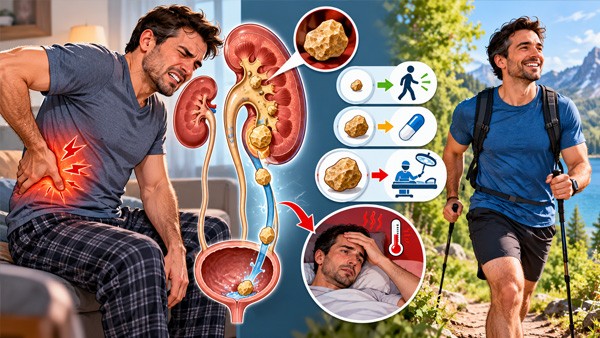
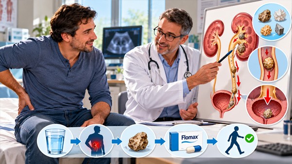
Renal colic is one of the few pains that people describe as the worst of their life, and one of the few where the right answer is usually to wait rather than to operate. Both of those are true at once, which is why the condition is so badly understood.
🪨 The five things that decide everything.
🔹 The pain is not the stone, it is the blockage. A stone sitting quietly in the kidney can be completely painless for years. The pain starts when it enters the ureter and the ureter cramps against an obstruction.
🔹 Size predicts whether it passes. Under 4 mm it almost always passes by itself. Over 10 mm it almost never does. This is measured, not guessed.
🔹 An anti-inflammatory works better than an opioid here, because it treats the mechanism rather than masking it.
🔹 Drinking more water does not push out a stone that is already stuck. Fluid prevents the next stone. It does not move this one, and forcing fluids against an obstruction makes the pain worse.
🔹 A stone plus a fever is an emergency. An obstructed kidney that is also infected turns into sepsis within hours, and antibiotics alone do not rescue it.
📏 Size, and what it actually predicts
This is the first question a urologist answers after the scan, because it decides between waiting and intervening.
| Stone size | Chance of passing without a procedure | Typical time |
|---|---|---|
| 1 to 2 mm | Very high — most people never know they had one | Days |
| 3 to 4 mm | Around 80 percent | One to two weeks |
| 5 to 6 mm | Roughly half | Two to four weeks |
| 7 to 9 mm | Low, and worth planning for a procedure | Often does not pass |
| 10 mm and above | Very unlikely | Intervention is the expected route |
Position matters almost as much as size. A stone in the lower third of the ureter, close to the bladder, has a far better chance than the same stone sitting high up, and most stones that are going to pass do so within four weeks. A stone that has not moved in six weeks is unlikely to move at all, and waiting past that point risks quiet damage to the kidney behind it.
🗾 Why the pain moves, and what the move means
The pain travels as the stone descends, and following it tells you roughly where things stand.
- 🔹 Flank, below the ribs at the back — the stone is at the top, where the kidney drains into the ureter
- 🔹 Pain wrapping round towards the front and down — it has entered the middle of the ureter
- 🔹 Groin, testicle or labia — the lower ureter. This is normal referred pain and not a separate problem
- 🔹 Constant urge to urinate, with burning and small volumes — the stone is at the junction with the bladder and is irritating it. This stage is frequently mistaken for a urinary infection, and it is the point at which the stone is closest to passing
- 🔹 Pain stops suddenly — usually the stone has entered the bladder. From the bladder out is painless, and most people never notice it leave
💡 Two things that surprise people.
🔹 Renal colic comes in waves and you cannot find a comfortable position. That restlessness is characteristic. Pain that is relieved by lying still is more likely to be something else, which is a genuinely useful distinction.
🔹 Nausea and vomiting are part of the picture, not a sign of food poisoning or a separate illness. The kidney and the gut share nerve supply, so severe colic produces vomiting in most people.
🚨 The combination that cannot wait
🚨 Go to hospital immediately, not in the morning, if any of these are present:
🔹 Fever or shaking chills with the pain. This is the single most important line on the page. A blocked kidney that is also infected is a closed infected space, and it progresses to sepsis in hours
🔹 Vomiting so persistent that you cannot keep fluids or tablets down
🔹 Pain that is not controlled by anything you have been given
🔹 Passing little or no urine, or pain on both sides at once
🔹 You have one working kidney, a transplanted kidney, or known kidney disease
🔹 Confusion, a racing pulse, clammy skin or feeling faint
An infected obstructed kidney is treated by draining it — a stent passed up from the bladder, or a tube through the back — together with antibiotics. The antibiotic cannot reach a space that has no outflow, which is the same reason an abscess anywhere has to be drained rather than medicated.
And one that is not about stones at all. A first episode of what looks like renal colic in someone over fifty, particularly with back pain, a history of smoking or high blood pressure, needs imaging rather than reassurance. A leaking abdominal aortic aneurysm produces flank pain radiating to the groin and has been repeatedly mistaken for a kidney stone. If you have had stones before and this feels identical, that is reassuring. If this is the first time and you are older, it is not.
💊 Pain control: why the anti-inflammatory wins
This is the part most often got wrong, including in hospitals.
- ✅ A non-steroidal anti-inflammatory is the first choice in renal colic, not a second-line option. It reduces the pressure inside the obstructed system and calms the ureteric smooth muscle, so it works on the cause of the pain rather than only the sensation
- 📉 It also causes less vomiting than opioids, which matters a great deal in a condition that already makes people sick
- 💊 Voltaren (Diclofenac) and Indocin (Indomethacin) are the agents with the longest record in renal colic specifically; Motrin (Ibuprofen) is the gentler everyday option
- 🏥 Opioids are added when the anti-inflammatory is not enough or cannot be used, rather than used first
- ⚠️ Anti-inflammatories are not for everyone. Existing kidney disease, dehydration, stomach ulcers, heart failure and certain other medicines change the calculation, and in a kidney that is already obstructed the decision belongs to a clinician. The general comparison is in which painkiller to take and who should avoid anti-inflammatories
- ⏰ Take it regularly for the first days rather than waiting for each wave. Colic that is already peaking is much harder to bring down
- 🌡️ Heat on the flank helps. A hot pack or a warm bath genuinely reduces colic pain and is one of the few home measures with real effect
🚪 What tamsulosin does, honestly
Alpha blockers relax the smooth muscle of the lower ureter, which is where most stones get stuck. Marketing around this has been enthusiastic and the evidence is more measured, so here is the real position.
📊 Where it helps and where it does not.
🔹 The benefit is concentrated in larger distal stones — roughly 5 to 10 mm in the lower third of the ureter. There it shortens passage time and reduces the number of pain episodes.
🔹 For small stones under 5 mm the benefit is minimal, because those were going to pass anyway. Large trials that mixed all sizes together found little overall effect, which is why the picture looks contradictory.
🔹 It is also used before and after procedures to reduce stent discomfort and help fragments clear.
🔹 Side effects are mild and specific: dizziness on standing, blocked nose, and retrograde ejaculation, which is harmless and reverses when the drug stops.
🔹 Using it this way is off-label in many countries. That is normal and widespread in urology, and it should be a decision made with a clinician rather than a self-started course.
💧 The water myth, taken apart
"Drink a lot of water and flush it out" is the most repeated piece of advice about kidney stones, and it mixes up two completely different things.
- ❌ Fluid does not push out a lodged stone. The stone is wedged in a tube roughly 3 mm wide. Increasing the flow behind it raises the pressure, which raises the pain, and does not widen the tube
- ⚠️ Forcing large volumes during an attack can make the colic worse, and in a fully obstructed kidney it is counterproductive
- ✅ Drink enough to stay comfortable and not become dehydrated, especially if you are vomiting. That is the goal during an attack, not flushing
- 💪 Fluid intake is the strongest single preventive measure there is — but between stones, not during one. The target is about 2.5 litres of urine a day, which usually means drinking around three litres
- 🏃 Movement is reasonable, walking around rather than lying rigid, though no particular exercise or position has been shown to dislodge a stone
🔬 Diagnosis, and the test result that misleads
| Test | What it does and does not settle |
|---|---|
| Low-dose CT without contrast | The reference test. Shows the stone, its exact size and position, and the degree of obstruction. Low-dose protocols cut the radiation substantially |
| Ultrasound | First choice in pregnancy, in children and in young adults. Shows obstruction well, measures stones less precisely, and misses small ones in the mid ureter |
| Urine dipstick | Blood is present in around 85 percent of cases, which means its absence does not rule a stone out. This is the result that most often sends people down the wrong path |
| Urine culture | Looks for infection alongside the stone, which changes urgency completely |
| Blood tests | Kidney function, calcium, and signs of infection |
| Plain x-ray | Of limited use on its own — uric acid stones are invisible on it |
And one practical instruction that people skip: strain your urine through a filter or fine sieve and keep what you catch. Knowing what the stone is made of changes the entire prevention plan, and the stone itself is the only way to know for certain. A stone passed into a toilet is information lost permanently.
🧪 What the stone is made of changes the plan
| Type | Share | What it tells you |
|---|---|---|
| Calcium oxalate | Around 70 to 80 percent | The default. Fluid, less sodium, normal calcium, moderate oxalate, citrate |
| Calcium phosphate | About 10 percent | Often linked to alkaline urine or a kidney tubule disorder, and worth investigating |
| Uric acid | Around 5 to 10 percent | The only common stone that can be dissolved rather than removed. Alkalinising the urine plus Zyloprim (Allopurinol) can shrink it. Strongly associated with gout — see why treating each gout attack never ends it |
| Struvite | A few percent | Infection stones. They grow fast, can fill the whole kidney, and must be removed completely, because any fragment left behind regrows the stone. Antibiotics do not clear them |
| Cystine | Rare | An inherited condition. Usually starts in childhood or early adulthood and needs specialist management |
🦴 The calcium mistake
✅ Cutting calcium out of your diet makes calcium stones more likely, not less.
🔹 Why: calcium in food binds oxalate in the gut and carries it out in the stool. Remove the calcium and the free oxalate is absorbed instead, reaches the kidney and forms stones there.
🔹 What to do: keep a normal calcium intake from food, around 1000 to 1200 mg a day, and take it in the same meal as the oxalate-rich food so the two meet in the gut rather than in the kidney.
🔹 Calcium supplements are different from calcium in food, and taken between meals they can raise risk. If they are needed for bone reasons, they go with food.
🔹 Sodium is the quiet driver. Salt makes the kidney excrete more calcium, so cutting salt lowers urinary calcium more reliably than cutting dairy ever did.
🥗 Diet, in order of how much it matters
- 💧 Fluid. Far ahead of everything else. Enough that urine stays pale through the day, including a glass at night if you wake
- 🧂 Less sodium. Mostly bread, processed meat, cheese, sauces and restaurant food rather than the salt shaker
- 🥩 Less animal protein. Large meat intake raises uric acid and urinary calcium and lowers citrate, which is three disadvantages from one habit
- 🍋 Citrate. Citrate blocks crystal formation directly. Lemon or lime juice in water is the practical version; prescribed potassium citrate is the stronger one
- 🥬 Oxalate, but only if your stones are oxalate. The heavy contributors are spinach, rhubarb, beetroot, almonds, peanuts, cocoa, black tea and wheat bran. Eating them with a source of calcium removes most of the risk, which is a far easier instruction than avoidance
- 🍬 Less sugary drink and less fructose, both independently associated with stones
- 🍊 Vitamin C supplements above about a gram a day raise oxalate and have been linked to stones. Vitamin C from food has not
💊 Medicines and conditions that make stones
Recurrent stones with no obvious dietary cause often have one of these behind them.
- 🔹 Topiramate and acetazolamide — used for epilepsy, migraine and glaucoma. Both alkalinise the urine and lower citrate, and both are a recognised cause
- 🔹 High-dose vitamin C, and calcium supplements taken away from food
- 🔹 Some antivirals and sulfadiazine, which crystallise in the urine directly
- 🔹 Primary hyperparathyroidism — the commonest missed cause. A raised blood calcium alongside stones should always be followed up, because the stones stop once the gland is dealt with
- 🔹 Gout and high uric acid
- 🔹 Inflammatory bowel disease, bowel resection and bariatric surgery, all of which raise oxalate absorption sharply
- 🔹 Type 2 diabetes, obesity and metabolic syndrome, which make the urine persistently acidic. Related: diabetic kidney disease and why it stays silent
- 🔹 Renal tubular acidosis and other inherited tubule disorders, usually presenting young and with calcium phosphate stones
- 🔹 Repeated urinary infections, which produce struvite stones — see what actually treats urinary tract infections
Thiazide diuretics such as Esidrix (Hydrochlorothiazide) sit on the other side of this list — they lower urinary calcium and are used deliberately to prevent recurrent calcium stones, which is one of the few places a blood pressure drug is prescribed for a kidney reason.
🔧 The procedures, and what each one is like
| Procedure | When, and what to expect |
|---|---|
| Shock wave lithotripsy | Shock waves from outside break the stone up. No incision, usually a day case. Best for smaller stones in the kidney or upper ureter. Fragments then have to pass, so colic afterwards is normal. Not used in pregnancy |
| Ureteroscopy with laser | A thin scope passed up through the bladder, the stone broken with a laser and the fragments removed. The highest clearance rate for ureteric stones, and usually leaves a temporary stent |
| Percutaneous nephrolithotomy | Through a small tract in the back, for large or branched kidney stones. The route for struvite stones filling the kidney. A short hospital stay |
| Stent alone | An urgent measure to relieve obstruction, especially with infection. It does not treat the stone, it buys safety until the stone can be dealt with |
💡 About stents, because nobody warns people. A ureteric stent commonly causes urgency, frequency, blood in the urine and a dragging ache in the flank when you urinate. That is expected and not a sign anything has gone wrong. It settles when the stent comes out, and an alpha blocker is often given specifically to reduce it. Fever with a stent in place, however, still means medical attention.
🔁 It is a recurring disease, not a one-off event
This is the framing that changes behaviour, and it is rarely stated clearly.
- 📈 Roughly half of people who form one stone form another within five to ten years without any preventive change
- 📉 Fluid alone roughly halves that, which is a larger effect than most prescription medicines achieve for anything
- 🔍 A metabolic workup is worth asking for after a second stone, after a first stone in a young person or a child, with stones on both sides, with a strong family history, or when the stone was not calcium oxalate. It is usually a 24-hour urine collection plus blood calcium and uric acid
- 📋 Prevention is then targeted — potassium citrate, a thiazide, or allopurinol, chosen by what the urine shows rather than by guesswork
- 🏥 Stones are not harmless between attacks. Repeated obstruction and repeated infection cause lasting loss of kidney function, and stone formers carry a measurably higher rate of chronic kidney disease — see chronic kidney disease, stages and the medicines that matter
😰 Flank pain that is not a stone
| Cause | What points to it instead |
|---|---|
| Kidney infection | Fever, chills, a tender back and feeling genuinely ill, with a more constant ache than colic. Needs antibiotics promptly |
| Leaking aortic aneurysm | First episode, older age, smoking or hypertension, sometimes a pulsing abdominal mass. The one that must not be assumed to be a stone |
| Musculoskeletal back pain | Worse on specific movements, better lying still. Colic does not let you keep still |
| Shingles before the rash appears | Burning pain in a band on one side, skin sensitive to clothing. The rash follows a few days later |
| Ovarian cyst or torsion, ectopic pregnancy | Lower abdominal rather than flank, and a pregnancy test is part of assessing any woman of childbearing age with this pain |
| Appendicitis, diverticulitis, biliary colic | Pain that localises to one quadrant and stays there, with tenderness to pressure |
| Bladder spasm or overactive bladder | Urgency and frequency without the flank component — see what calms bladder spasms |
🤰 Stones in pregnancy
- 🔹 Ultrasound first, always. CT is avoided, and low-dose magnetic resonance imaging is the next step if ultrasound is not enough
- 🔹 Most pass with fluids and pain relief, as outside pregnancy
- 🔹 Anti-inflammatories are generally avoided, particularly after the middle of pregnancy, so pain control looks different and is managed by the obstetric team
- 🔹 Shock wave lithotripsy is not used. When intervention is needed it is a stent or ureteroscopy
- 🔹 Infection is taken more seriously, because an obstructed infected kidney in pregnancy carries risk for both the pregnancy and the mother
- 🔹 A stent or nephrostomy placed in pregnancy needs changing more often, because it encrusts faster
👶 Stones in children
Stones in a child are uncommon enough that they always deserve investigation rather than simple treatment. The presentation is often vague — abdominal pain, vomiting, blood in the urine, or recurrent urinary infection rather than classic colic. Ultrasound is the first test, and a full metabolic assessment is standard, because an inherited cause or a tubule disorder is much more likely than in an adult.
📞 When to seek help, and how fast
🚨 Emergency, now: fever or chills with the pain; vomiting that stops you keeping anything down; pain that nothing controls; little or no urine; pain on both sides; one kidney, a transplant or known kidney disease; confusion, faintness or a racing pulse; a first episode of this pain over the age of fifty.
Same day:
- Visible blood in the urine, whether or not there is pain
- Pain that has settled but keeps returning in waves over several days
- Pregnancy with flank pain
Arrange a review if:
- Four weeks have passed and the stone has not come out — the kidney behind it needs checking even if the pain has eased
- You have had two or more stones, and have never had a metabolic workup
- You caught the stone — take it in for analysis
- You take topiramate, acetazolamide, high-dose vitamin C or calcium supplements and have formed a stone
- Your blood calcium has been reported as high at any point
- You get recurrent urinary infections alongside stones
❓ Frequently asked questions
How long does it take to pass a kidney stone?
Small stones under 4 mm usually pass within one to two weeks. Stones of 5 to 6 mm may take four weeks. A stone that has not moved in six weeks is unlikely to pass.
Does drinking lots of water flush out a kidney stone?
No. Extra fluid does not move a stone that is already wedged, and forcing it during an attack can worsen the pain. Fluid prevents the next stone rather than passing this one.
When is a kidney stone an emergency?
Fever or chills with the pain is the key sign, because an obstructed infected kidney becomes sepsis within hours. Also uncontrolled pain, persistent vomiting, or passing no urine.
What is the best painkiller for kidney stone pain?
An anti-inflammatory such as diclofenac or indomethacin is first choice, because it lowers pressure in the ureter rather than only masking pain, and causes less vomiting than opioids.
Does tamsulosin help a stone pass?
Mainly for larger stones of 5 to 10 mm in the lower ureter, where it shortens passage time. For stones under 5 mm the benefit is small, since those usually pass regardless.
Should I cut out calcium to prevent kidney stones?
No, that makes them more likely. Dietary calcium binds oxalate in the gut so it leaves in the stool. Keep calcium normal and eat it with oxalate-rich food. Cut salt instead.
Can I have a kidney stone without blood in my urine?
Yes. Blood is found in about 85 percent of cases, so a clear dipstick does not rule out a stone. Imaging settles it, not the urine test.
Why do I need to urinate constantly with a kidney stone?
The stone has reached the junction with the bladder and is irritating it. It mimics a urinary infection, and it is the stage at which the stone is closest to passing.
Will I get another kidney stone?
About half of stone formers do within five to ten years with no preventive change. Raising fluid intake roughly halves that, and targeted prevention lowers it further.
Why should I keep the stone after it passes?
Its composition decides the prevention plan, and the stone is the only certain way to know. Uric acid stones can be dissolved; struvite stones mean infection needs addressing.
📑 Sources and editorial
- Urological guidance on the management of urolithiasis, including stone size and location as predictors of spontaneous passage, the four to six week observation window, and indications for intervention
- Guidance on acute renal colic, including non-steroidal anti-inflammatory drugs as first-line analgesia, the comparison with opioids for pain relief and vomiting, and the physiological basis for their effect on intraureteric pressure
- Trial and meta-analytic evidence on medical expulsive therapy with alpha blockers, including the concentration of benefit in distal ureteric stones above 5 mm and the absence of meaningful effect in smaller stones
- Guidance on emergency management of the obstructed infected kidney, including the requirement for urgent decompression by ureteric stent or percutaneous nephrostomy in addition to antimicrobial therapy
- Literature on the misdiagnosis of ruptured abdominal aortic aneurysm as renal colic, and the recommendation to image a first presentation of flank pain in older patients
- Diagnostic guidance on low-dose non-contrast computed tomography as the reference standard, ultrasound as first-line in pregnancy and in younger patients, and the reported sensitivity of urinary dipstick haematuria
- Data on stone composition frequencies, including the predominance of calcium oxalate, and the dissolution of uric acid stones by urinary alkalinisation
- Guidance on struvite and infection-related stones, including the requirement for complete clearance to prevent regrowth
- Evidence on dietary calcium and stone risk, including the inverse relationship between dietary calcium intake and calcium oxalate stone formation, and the contrasting effect of supplements taken apart from meals
- Evidence on sodium intake and urinary calcium excretion, animal protein and urinary citrate, and the association between sugar-sweetened beverage intake and stone formation
- Evidence on high-dose ascorbic acid supplementation and urinary oxalate excretion
- Guidance on preventive pharmacotherapy, including thiazide diuretics for hypercalciuria, alkali citrate for hypocitraturia, and allopurinol for hyperuricosuric stone formation
- Literature on drug-induced urolithiasis, including topiramate, acetazolamide, sulfadiazine and protease inhibitors
- Guidance on secondary causes of stone disease, including primary hyperparathyroidism, enteric hyperoxaluria after bowel resection or bariatric surgery, renal tubular acidosis and cystinuria
- Evidence on stone recurrence rates and on the reduction achieved by increased fluid intake, and indications for metabolic evaluation
- Evidence on the association between a history of nephrolithiasis and subsequent chronic kidney disease
- Guidance on shock wave lithotripsy, ureteroscopy with laser lithotripsy and percutaneous nephrolithotomy, including stone-free rates and selection criteria
- Literature on ureteric stent-related symptoms and the use of alpha blockers to reduce them
- Guidance on the management of urolithiasis in pregnancy, including imaging selection, the contraindication to shock wave lithotripsy, and more frequent stent exchange
- Guidance on paediatric urolithiasis, including the atypical presentation and the standard use of full metabolic evaluation
- Related reading: what actually treats urinary tract infections, recurrent cystitis and when prophylaxis is justified, bladder spasms and urgency, benign prostatic hyperplasia and which drug works in days, chronic kidney disease stages, gout and the uric acid target, which painkiller to take
- Related products: Flomax (Tamsulosin), Voltaren (Diclofenac), Indocin (Indomethacin), Motrin (Ibuprofen), Zyloprim (Allopurinol), Esidrix (Hydrochlorothiazide), Cipro (Ciprofloxacin)
- RXshop Editorial Team — reviewed by Michael Reeves, MD, FACS — Urologist
Medical Disclaimer: The information in this article is for educational and informational purposes only and is not a substitute for professional medical advice, diagnosis, or treatment. Always seek guidance from a qualified healthcare provider with any questions you may have regarding a medical condition, and before starting, stopping or changing any medication.